Home
/ Bone Cross Section Diagram Labeled - Bone Cross Section High Res Stock Images Shutterstock - Compact bone is the denser, stronger of the two types of bone tissue ( (figure) ).
Bone Cross Section Diagram Labeled - Bone Cross Section High Res Stock Images Shutterstock - Compact bone is the denser, stronger of the two types of bone tissue ( (figure) ).
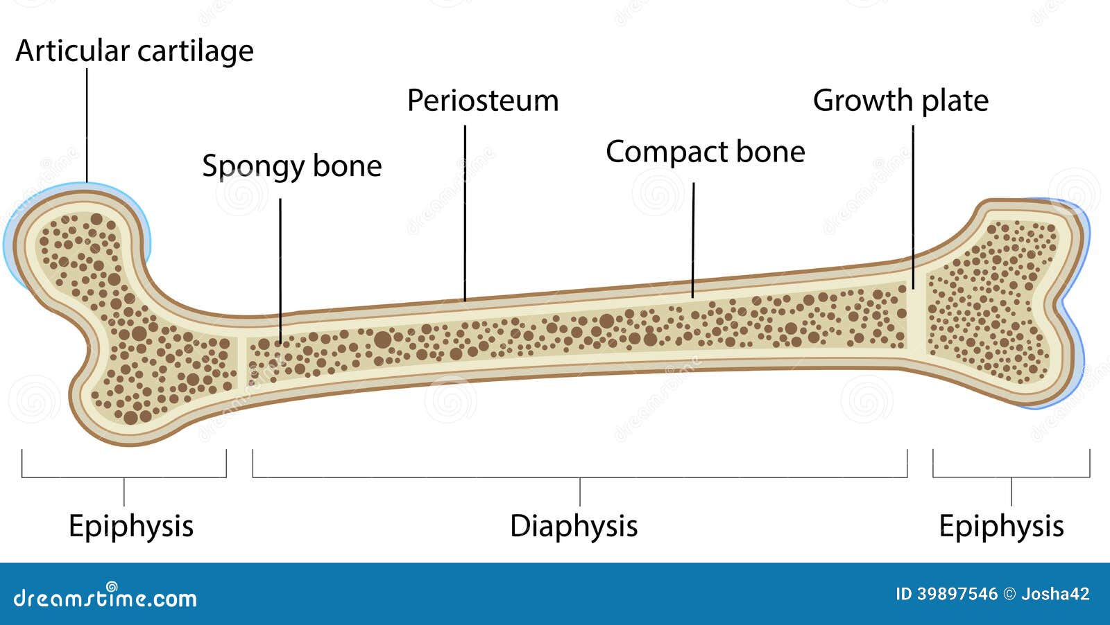
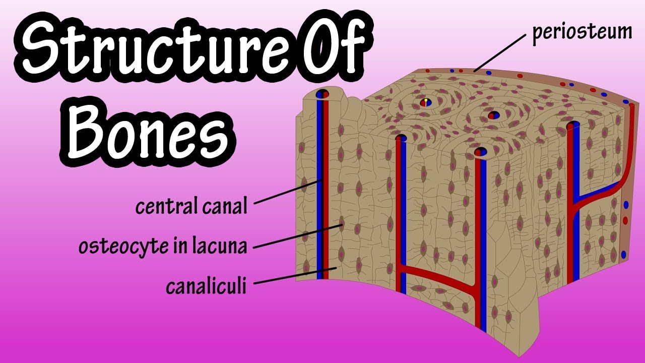
Bone Cross Section Diagram Labeled - Bone Cross Section High Res Stock Images Shutterstock - Compact bone is the denser, stronger of the two types of bone tissue ( (figure) ).. Storage of fat for a ready energy source for active muscles. Get express international delivery to your door. The muscle acts as the effort force; A cross section of a human long bone. The osteocytes are arranged in concentric rings of bone matrix called lamellae (little plates), and their processes run in interconnecting canaliculi.
Get express international delivery to your door. For example, to read this diagram literally, since the cartilage can be seen inside the cutaway section of bone, it incorrectly indicates that the cartilage in fact goes through the bone structure, rather than just being found around the bone end. This is a cross section through decalcified bone. The diaphysis is the tubular shaft that runs between the proximal and distal ends of the bone. Fixed slide cross section of muscle tissue, 100x microscope view.
Bone Anatomy Labeled Diagram Stock Vector Illustration Of Femur Medicine 39897546 from thumbs.dreamstime.com The periosteum contains many strong collagen fibers that are used to firmly anchor tendons and muscles to the bone for movement. There are two ways to study bone histology. Diagram orienting yourself within such a cross section is easy. The central haversian canal, and horizontal canals (perforating/ volkmann's) canals contain blood vessels and nerves from the periosteum. The point of attachment on the more movable bone in the illustration is called the _____ of the muscle. This is a cross section through decalcified bone. Red marrow fills the spaces in some bones. Cross section of human bone diagram 12 photos of the cross section of human bone diagram cross section diagram of human bone, bone, cross section diagram of human bone.
Download 706 bone cross medical section stock illustrations, vectors & clipart for free or amazingly low rates!
Browse 4,294 bone cross section stock photos and images available, or search for human bone cross section to find more great stock photos and pictures. The periosteum contains many strong collagen fibers that are used to firmly anchor tendons and muscles to the bone for movement. A long bone has two parts: Related posts of cross section of human bone diagram bone in arm pictures. It seems confusing and misleading. Diagram orienting yourself within such a cross section is easy. Bones in your body names. Bodytomy provides a labeled diagram of the haversian system to help you the terms 'haversian system' or 'osteon' refer to the basic. Damage in even one part can hinder the functioning of the knee. The diagram above shows a transverse view. Find the perfect bone cross section stock photos and editorial news pictures from getty images. Cross section of a long bone. Cross section of a bone, this image shows the interior of the bone, which has a lot of spongy bone tissue.
Cross section of a bone. Which labeled structure in the given image is a fascicle? I don't like way you've shown the cartilage. The point of attachment on the more movable bone in the illustration is called the _____ of the muscle. New users enjoy 60% off.
Bone Cross Section Diagram Labeled 19 The Respiratory System Medicine Libretexts Cartilage And Bone Are Flexible And Rigid Connective Tissue from i1.wp.com The structure of a long bone allows for the best visualization of all of the parts of a bone ( figure 6.7 ). Related posts of bone cross section labeled. For example, to read this diagram literally, since the cartilage can be seen inside the cutaway section of bone, it incorrectly indicates that the cartilage in fact goes through the bone structure, rather than just being found around the bone end. From which part of the sarcomere is the given cross section taken? The star of the show (brain) is easily recognizable because it appears highly convoluted, full of ridges (gyri) and indentations (sulci).the paired thalami appear as two circular masses in the midline, forming the walls of the third ventricle.the neurocranium appears as a meshwork (trabecular. It can be found under the periosteum and in the diaphyses of long bones, where it provides support and protection. I don't like way you've shown the cartilage. Click to edit this example.
The structure of a long bone allows for the best visualization of all of the parts of a bone ( figure 6.7 ).
Save on your favourite brand labels today with saks off 5th. The central haversian canal, and horizontal canals (perforating/ volkmann's) canals contain blood vessels and nerves from the periosteum. Download 706 bone cross medical section stock illustrations, vectors & clipart for free or amazingly low rates! The diaphysis is the tubular shaft that runs between the proximal and distal ends of the bone. At a symphysis, the bones are joined by fibrocartilage. Jump to navigation jump to search. Looking at a bone in cross section, there are several distinct layered regions that make up a bone. Cross section of a bone, this image shows the interior of the bone, which has a lot of spongy bone tissue. There are trabeculae in spongy bone which gives its sponge like appearance. Anatomy of a flat bone. From which part of the sarcomere is the given cross section taken? For example, to read this diagram literally, since the cartilage can be seen inside the cutaway section of bone, it incorrectly indicates that the cartilage in fact goes through the bone structure, rather than just being found around the bone end. Get express international delivery to your door.
It seems confusing and misleading. Find the perfect bone cross section stock photos and editorial news pictures from getty images. Related posts of cross section of human bone diagram bone in arm pictures. Which labeled structure in the given image is a fascicle? From which part of the sarcomere is the given cross section taken?
Structure Of Bone Tissue Bone Structure Anatomy Components Of Bones Youtube from i.ytimg.com A long bone has two parts: Save on your favourite brand labels today with saks off 5th. Smartdraw includes 1000s of professional healthcare and anatomy chart templates that you can modify and make your own. At a symphysis, the bones are joined by fibrocartilage. Damage in even one part can hinder the functioning of the knee. The star of the show (brain) is easily recognizable because it appears highly convoluted, full of ridges (gyri) and indentations (sulci).the paired thalami appear as two circular masses in the midline, forming the walls of the third ventricle.the neurocranium appears as a meshwork (trabecular. There are trabeculae in spongy bone which gives its sponge like appearance. Compact bone is the denser, stronger of the two types of bone tissue ( (figure) ).
Cross section of a bone, this image shows the interior of the bone, which has a lot of spongy bone tissue.
Indicated are haversian canal with blood and lymphatic vessels, a nerve, and loose. The diagram above shows a transverse view. Bodytomy provides a labeled diagram of the haversian system to help you the terms 'haversian system' or 'osteon' refer to the basic. The compact bone is made up of osteon. Human skeleton anatomy human body anatomy human anatomy and physiology muscle anatomy hand bone anatomy anatomy bones the human body anatomy images anatomy practice. A long bone has two parts: The diaphysis and the epiphysis. Bone matrix and cells bone matrix osseous tissue is a connective tissue and like all connective tissues contains relatively few cells and large amounts of extracellular matrix. Related posts of cross section of human bone diagram bone in arm pictures. Download 706 bone cross medical section stock illustrations, vectors & clipart for free or amazingly low rates! Cross section of a bone, this image shows the interior of the bone, which has a lot of spongy bone tissue. The periosteum contains many strong collagen fibers that are used to firmly anchor tendons and muscles to the bone for movement. There are two ways to study bone histology.
Bone decalcification is the removal of the mineral component using an acid, leaving the bone soft and easy to cut bone cross section. A long bone has two parts:

 ).)